
Same-day urgent eye care
Mon 10–5 · Tue–Fri 8–5 310-828-2010 Book online Intake form
Cohenmehr Optometry — Vision · Dry Eye · Keratoconus Book appointment
Cohenmehr OptometryVision · Dry Eye · Keratoconus Center
Our Practice
Meet the Doctors TestimonialsShare FeedbackTraveling to See UsAreas We Serve Hours & Directions
Eye Care

Routine care

Comprehensive Eye CareContact Lens ExamsComputer Vision SyndromeGlasses and Contact LensesNeurolensAdvanced Diagnostics

Medical eye care

Glaucoma & Ocular DiseaseDiabetic Eye ExamsMacular Degeneration CareLow VisionStye & Chalazion TreatmentUrgent Care

Children’s vision

Pediatric Eye ExamsMyopia ManagementAtropineOrtho-KEssilor Stellest LensesVision Therapy

Surgery co-management

Surgical Co-ManagementLASIKPRKSMILEEVO ICLRefractive Lens ExchangeCataract SurgeryCorneal TransplantPterygium Surgery
Dry Eye
The Dry Eye CenterOptiLIFTIPL (Intense Pulsed Light)Punctal PlugsBlephExAmniotic MembranesPrescription DropsAutologous Serum DropsMeibomian Gland ProbingIPL for Rosacea

Understand

Types of Dry EyeThe Tear FilmDemodex BlepharitisDry Eye After LASIKMenopause & Dry EyeSjögren’s & Dry EyeContact Lens Discomfort

Cosmetics

Upneeq® · Buy OnlineUpneeq Intake Form
Myopia Control
The Myopia ProgramMyopia Estimator — free toolOrtho-K — $1,500 totalAtropineStellest LensesMultifocal ContactsMiSight 1 Day

For parents

Choosing a TreatmentWhy Myopia MattersOutdoor Time & ScreensFirst Ortho-K NightMeasuring Progression
Keratoconus
The Keratoconus CenterScleral Contact LensesSpecialty Contact LensesScleral Lens UsesEaglet Surface ProfilerScleral Lens Care

Go deeper

Eye Rubbing & KeratoconusKeratoconus in TeensCorneal HydropsTransplant Co-Management
Scleral Lenses

The lenses

Scleral Contact LensesEvery Use, at a GlanceEaglet Surface ProfilerScleral vs RGP vs HybridSpecialty Contact LensesSpecialty Soft & ProstheticLens Care & HandlingTraveling to See Us

Corneal conditions

KeratoconusPost-LASIK EctasiaPellucid Marginal DegenerationCorneal ScarringAfter Corneal TransplantAfter Radial Keratotomy (RK)Post-Surgical ComplicationsOcular Shingles Scarring

Surface disease & more

Severe Dry EyeSjögren’s SyndromeStevens-Johnson SyndromeGraft-vs-Host DiseaseBell’s Palsy & ExposureRare Tear DeficiencyPresbyopia & High RxSports & Active Wear
Library
Start With a SymptomEye Conditions A–ZArticlesFree ToolsFull Site Index

Patients

Patient Intake FormCommon QuestionsAccepted InsurancePaying for Care (FSA/HSA)
Call the office310-828-2010
Book appointment

Medically reviewed by Dr. Kevin Cohenmehr, OD Last reviewed

Diabetic Retinopathy: Symptoms, Causes & Treatment

High blood sugar, small vessels, silent damage.

Diabetic retinopathy is what prolonged high blood sugar does to the retina’s small blood vessels: they leak, swell and close off. It is a leading cause of vision loss in working-age adults — and in its treatable early stages it usually causes no symptoms at all.
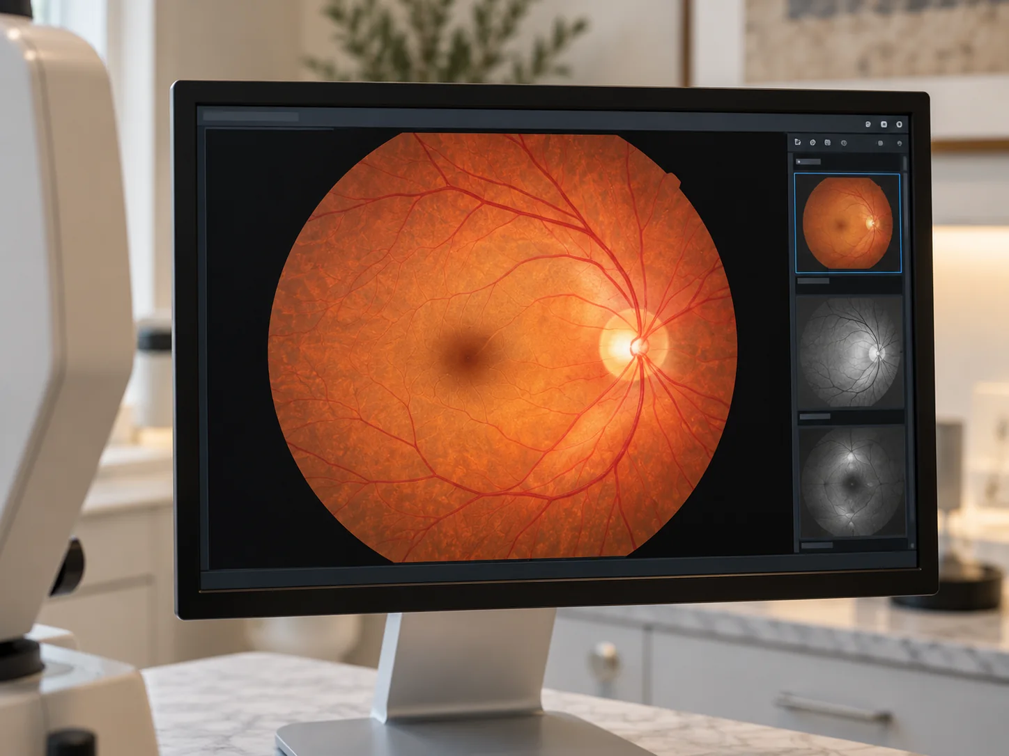
A retinal photograph showing the vessels
Cause
Vessel damage from diabetes
Early symptoms
Usually none
The defense
Annual dilated exams
Led by
Dr. Kevin Cohenmehr

Later-stage warning signs

  • New floaters or dark strands
  • Blurred or fluctuating vision
  • Dark or empty patches in the field of view
  • Trouble reading that glasses don’t fix

Don’t wait for symptoms

By the time retinopathy is noticeable, it is advanced. The annual dilated exam exists precisely because the early, treatable stages are silent.

Care

Caught early, managed together.
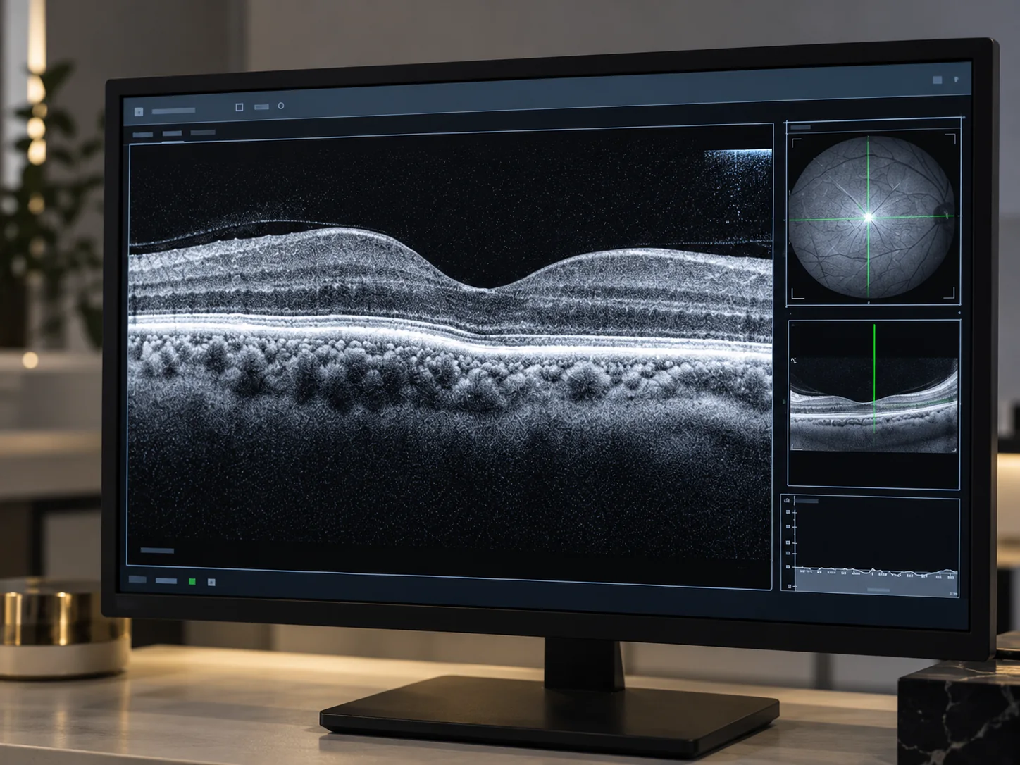
OCT imaging of the retina
  • Dilated retinal examinations with retinal photography for baseline and progression tracking
  • OCT imaging to detect swelling (macular edema), thinning or structural change
  • Findings reported to your physician or endocrinologist, because blood sugar control is the other half of treatment
  • Prompt referral to retina specialists for injections or laser when the stage calls for it

The schedule

Yearly exams protect working vision.

  • Every year for most people with diabetes
  • More often once changes appear
  • Same-day visits for sudden floaters or vision change
Book a diabetic eye exam

Common questions

Diabetic retinopathy, answered.

Anything else, call the office at 310-828-2010.

  • Soon after diagnosis for type 2, and on the schedule your doctors set from there — usually yearly.

  • Early changes can stabilize with blood sugar control; later stages are treated with injections or laser by retina specialists. Either way, early detection drives the outcome.

  • No — it lowers risk but doesn’t remove it. The yearly exam stays.

  • In the early, most treatable stages, none. Later: blurred vision that comes and goes with blood sugar, trouble reading, dark floating spots or cobweb streaks when fragile vessels bleed, and a blurred or empty area in the center of vision when the macula swells. Waiting for symptoms means waiting for damage, which is the case for the yearly dilated diabetic exam.

  • Non-proliferative retinopathy (mild to severe) is the early phase: small vessels leak, swell and close off. Proliferative retinopathy is the advanced phase: the starved retina grows fragile new vessels that bleed and scar. Diabetic macular edema, swelling of the central retina, can occur at any stage and is the usual cause of blurred vision. Each stage sets its own follow-up interval, as often as every two to four months in later stages.

Also consider

Next step

Protect your retina this year.

Book the annual exam with a Los Angeles optometrist — your physician gets the report, and you get certainty.

Book Today Or call 310-828-2010