
Same-day urgent eye care
Mon 10–5 · Tue–Fri 8–5 310-828-2010 Book online Intake form
Cohenmehr Optometry — Vision · Dry Eye · Keratoconus Book appointment
Cohenmehr OptometryVision · Dry Eye · Keratoconus Center
Our Practice
Meet the Doctors TestimonialsShare FeedbackTraveling to See UsAreas We Serve Hours & Directions
Eye Care

Routine care

Comprehensive Eye CareContact Lens ExamsComputer Vision SyndromeGlasses and Contact LensesNeurolensAdvanced Diagnostics

Medical eye care

Glaucoma & Ocular DiseaseDiabetic Eye ExamsMacular Degeneration CareLow VisionStye & Chalazion TreatmentUrgent Care

Children’s vision

Pediatric Eye ExamsMyopia ManagementAtropineOrtho-KEssilor Stellest LensesVision Therapy

Surgery co-management

Surgical Co-ManagementLASIKPRKSMILEEVO ICLRefractive Lens ExchangeCataract SurgeryCorneal TransplantPterygium Surgery
Dry Eye
The Dry Eye CenterOptiLIFTIPL (Intense Pulsed Light)Punctal PlugsBlephExAmniotic MembranesPrescription DropsAutologous Serum DropsMeibomian Gland ProbingIPL for Rosacea

Understand

Types of Dry EyeThe Tear FilmDemodex BlepharitisDry Eye After LASIKMenopause & Dry EyeSjögren’s & Dry EyeContact Lens Discomfort

Cosmetics

Upneeq® · Buy OnlineUpneeq Intake Form
Myopia Control
The Myopia ProgramMyopia Estimator — free toolOrtho-K — $1,500 totalAtropineStellest LensesMultifocal ContactsMiSight 1 Day

For parents

Choosing a TreatmentWhy Myopia MattersOutdoor Time & ScreensFirst Ortho-K NightMeasuring Progression
Keratoconus
The Keratoconus CenterScleral Contact LensesSpecialty Contact LensesScleral Lens UsesEaglet Surface ProfilerScleral Lens Care

Go deeper

Eye Rubbing & KeratoconusKeratoconus in TeensCorneal HydropsTransplant Co-Management
Scleral Lenses

The lenses

Scleral Contact LensesEvery Use, at a GlanceEaglet Surface ProfilerScleral vs RGP vs HybridSpecialty Contact LensesSpecialty Soft & ProstheticLens Care & HandlingTraveling to See Us

Corneal conditions

KeratoconusPost-LASIK EctasiaPellucid Marginal DegenerationCorneal ScarringAfter Corneal TransplantAfter Radial Keratotomy (RK)Post-Surgical ComplicationsOcular Shingles Scarring

Surface disease & more

Severe Dry EyeSjögren’s SyndromeStevens-Johnson SyndromeGraft-vs-Host DiseaseBell’s Palsy & ExposureRare Tear DeficiencyPresbyopia & High RxSports & Active Wear
Library
Start With a SymptomEye Conditions A–ZArticlesFree ToolsFull Site Index

Patients

Patient Intake FormCommon QuestionsAccepted InsurancePaying for Care (FSA/HSA)
Call the office310-828-2010
Book appointment

Medically reviewed by Dr. Brandon Cohenmehr, OD Last reviewed

Post-LASIK Ectasia: Symptoms, Causes & Treatment

When vision drifts years after surgery.

A small number of LASIK patients develop ectasia: the treated cornea weakens and begins to bulge, like keratoconus arriving after surgery. Glasses stop feeling crisp, night vision fills with halos, and prescriptions keep changing. Imaging confirms it; real options follow.
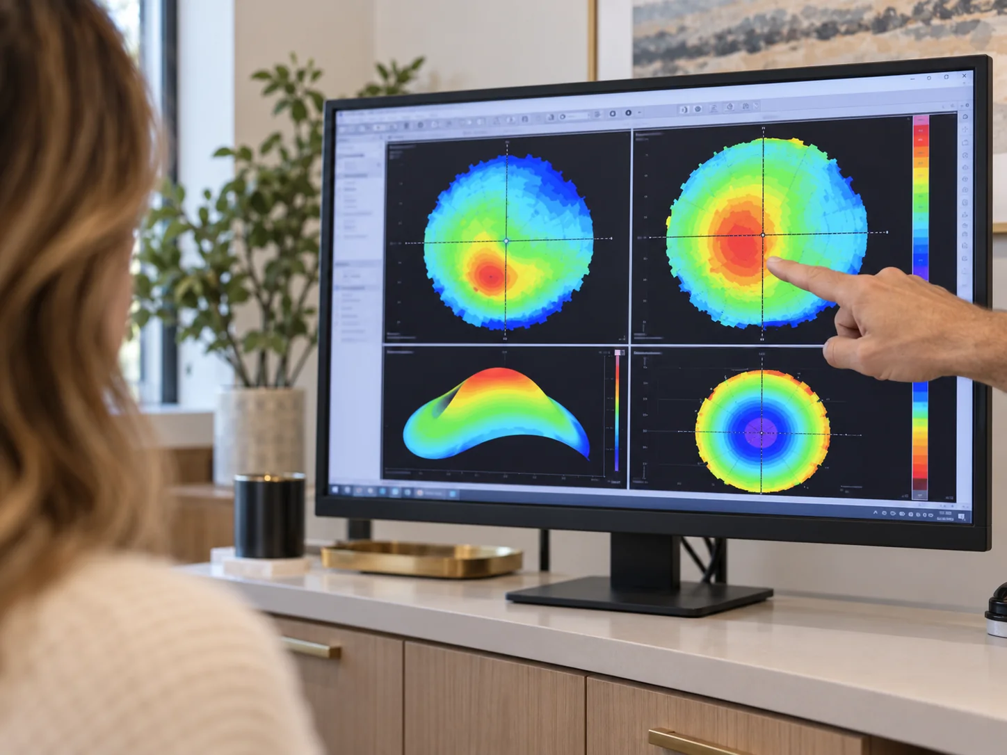
Corneal maps compared over time
What it is
Progressive corneal weakening
Signs
Ghosting, halos, shifting Rx
Confirmed by
Topography and tomography
Managed with
CXL + scleral lenses

Worth imaging if, after LASIK, you notice

  • Vision that keeps worsening despite new glasses
  • Ghost images or doubling in one eye
  • Halos and starbursts growing at night
  • One eye clearly weaker than the other
  • Astigmatism increasing at each exam

Two-part answer

Cross-linking (co-managed with corneal surgeons) can stabilize a progressing cornea; scleral lenses then restore the sharp vision glasses can’t. Protection first, optics second.

The pathway

Confirm, stabilize, see.

  • Corneal topography and tomography to confirm ectasia and set a baseline
  • Monitoring for progression — the decision driver
  • Referral for corneal cross-linking when the maps show change, with follow-up back here
  • Scleral lens fitting for vision: a smooth optical surface over the irregular cornea, designed from profilometry

Not your fault

Ectasia is rare, and it is manageable.

  • Modern screening has made it rarer; it still occasionally appears years later
  • Vision changing after LASIK deserves corneal imaging, not just stronger glasses
  • Most patients regain sharp functional vision with scleral lenses
Book a corneal evaluation

Common questions

Post-LASIK ectasia, answered.

Anything else, call the office at 310-828-2010.

  • It behaves like it — thinning and bulging — but appears after refractive surgery. Management is nearly identical.

  • The shape change isn’t reversed; cross-linking stabilizes it and scleral lenses correct the optics. Together they restore function.

  • Only imaging can say. That’s a routine part of a corneal evaluation here.

  • Rare. In a 2018 review of more than 30,000 LASIK-treated eyes followed for up to 8 years, 10 developed ectasia, about 0.03%. Most of those eyes had warning signs before surgery, such as a thin cornea or an irregular corneal map, which is why careful screening matters and why vision that keeps changing after LASIK deserves topography.

  • LASIK corrects vision by removing corneal tissue under a thin flap, which leaves the cornea thinner. When the cornea was weak to begin with (thin, or with early keratoconus that went undetected), or when too little untouched tissue remains after treatment, it can slowly bulge forward months or years later. The bulge does not reverse, but cross-linking can stabilize it and scleral lenses restore sharp vision.

Articles

By Dr. Brandon Cohenmehr, OD

Also consider

Next step

Changing vision after LASIK? Map it.

Book a corneal evaluation with topography and get a definitive answer from our optometrists in Los Angeles.

Book Today Or call 310-828-2010