Same-day urgent eye care
Mon 10–5 · Tue–Fri 8–5 310-828-2010 Book online Intake form
Cohenmehr Optometry — Vision · Dry Eye · Keratoconus Book appointment
Cohenmehr OptometryVision · Dry Eye · Keratoconus Center
Our Practice
Meet the Doctors TestimonialsShare FeedbackTraveling to See UsAreas We Serve Hours & Directions
Eye Care

Routine care

Comprehensive Eye CareContact Lens ExamsComputer Vision SyndromeGlasses and Contact LensesNeurolensAdvanced Diagnostics

Medical eye care

Glaucoma & Ocular DiseaseDiabetic Eye ExamsMacular Degeneration CareLow VisionStye & Chalazion TreatmentUrgent Care

Children’s vision

Pediatric Eye ExamsMyopia ManagementAtropineOrtho-KEssilor Stellest LensesVision Therapy

Surgery co-management

Surgical Co-ManagementLASIKPRKSMILEEVO ICLRefractive Lens ExchangeCataract SurgeryCorneal TransplantPterygium Surgery
Dry Eye
The Dry Eye CenterOptiLIFTIPL (Intense Pulsed Light)Punctal PlugsBlephExAmniotic MembranesPrescription DropsAutologous Serum DropsMeibomian Gland ProbingIPL for Rosacea

Understand

Types of Dry EyeThe Tear FilmDemodex BlepharitisDry Eye After LASIKMenopause & Dry EyeSjögren’s & Dry EyeContact Lens Discomfort

Cosmetics

Upneeq® · Buy OnlineUpneeq Intake Form
Myopia Control
The Myopia ProgramMyopia Estimator — free toolOrtho-K — $1,500 totalAtropineStellest LensesMultifocal ContactsMiSight 1 Day

For parents

Choosing a TreatmentWhy Myopia MattersOutdoor Time & ScreensFirst Ortho-K NightMeasuring Progression
Keratoconus
The Keratoconus CenterScleral Contact LensesSpecialty Contact LensesScleral Lens UsesEaglet Surface ProfilerScleral Lens Care

Go deeper

Eye Rubbing & KeratoconusKeratoconus in TeensCorneal HydropsTransplant Co-Management
Scleral Lenses

The lenses

Scleral Contact LensesEvery Use, at a GlanceEaglet Surface ProfilerScleral vs RGP vs HybridSpecialty Contact LensesSpecialty Soft & ProstheticLens Care & HandlingTraveling to See Us

Corneal conditions

KeratoconusPost-LASIK EctasiaPellucid Marginal DegenerationCorneal ScarringAfter Corneal TransplantAfter Radial Keratotomy (RK)Post-Surgical ComplicationsOcular Shingles Scarring

Surface disease & more

Severe Dry EyeSjögren’s SyndromeStevens-Johnson SyndromeGraft-vs-Host DiseaseBell’s Palsy & ExposureRare Tear DeficiencyPresbyopia & High RxSports & Active Wear
Library
Start With a SymptomEye Conditions A–ZArticlesFree ToolsFull Site Index

Patients

Patient Intake FormCommon QuestionsAccepted InsurancePaying for Care (FSA/HSA)
Call the office310-828-2010
Book appointment

Medically reviewed by Dr. Brandon Cohenmehr, OD Last reviewed

Pellucid Marginal Degeneration: Symptoms, Causes & Treatment

Keratoconus’s lesser-known cousin.

Pellucid marginal degeneration (PMD) thins a crescent along the lower edge of the cornea, letting the surface above it sag into a distinctive shape. Like keratoconus it distorts vision beyond what glasses can fix; unlike keratoconus it hides at the edge, where only good imaging looks.

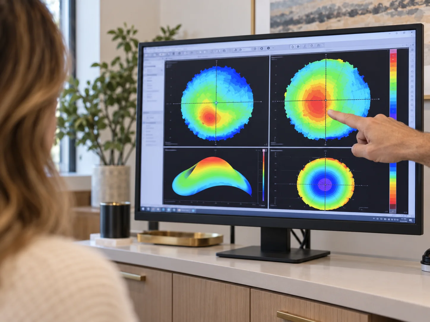
A corneal map with inferior steepening
Where
Thinning at the lower corneal edge
Signature
High against-the-rule astigmatism
Confirmed by
Tomography and pachymetry maps
Vision via
Scleral and specialty lenses

Care

Found at the edge, managed like a cone.

Diagnosis leans on tomography and thickness mapping, which catch the peripheral thinning a routine exam can miss. Management mirrors keratoconus: monitor for progression, consider cross-linking referral when it moves, and fit scleral lenses (which vault the entire irregular zone) for the vision glasses can’t deliver.

Because the thinning sits low, lens design matters even more; free-form scleral designs built from profilometry handle the asymmetry well.

Same toolkit

The keratoconus playbook applies.

Common questions

Pellucid marginal degeneration, answered.

Anything else, call the office at 310-828-2010.

  • The thinning sits at the lower margin rather than centrally, producing a distinctive map and heavy against-the-rule astigmatism. Treatment paths are nearly identical.

  • PMD hides at the corneal edge; it takes tomography — not just a glasses check — to see it.

  • Early on, partially. As the irregularity grows, scleral lenses become the reliable route to sharp vision.

  • Pellucid marginal degeneration (PMD) is a rare condition in which a band of the lower cornea near its edge, roughly from the 4 o'clock to the 8 o'clock position, slowly thins. The cornea just above the thinnest point bulges, causing astigmatism (uneven focusing) and blurrier vision, while the center of the cornea keeps its normal thickness. PMD affects both eyes, causes no inflammation and progresses slowly. The thinned area stays clear, which fits the name: "pellucid" means transparent.

  • PMD is rare, and it affects both eyes; it is a lesser-known relative of keratoconus that thins the lower edge of the cornea instead of its center. Because the thinning sits low at the edge, PMD is easy to miss: it shows up as astigmatism, and only tomography that maps the corneal edge reveals the thinning, which a routine glasses check does not. If your astigmatism keeps getting harder to correct with glasses, a corneal map is the way to check.

Also consider

Next step

Astigmatism that defies glasses?

Book a corneal evaluation with tomography at our eye clinic in Los Angeles — edges included.

Book Today Or call 310-828-2010