Same-day urgent eye care
Mon 10–5 · Tue–Fri 8–5 310-828-2010 Book online Intake form
Cohenmehr Optometry — Vision · Dry Eye · Keratoconus Book appointment
Cohenmehr OptometryVision · Dry Eye · Keratoconus Center
Our Practice
Meet the Doctors TestimonialsShare FeedbackTraveling to See UsAreas We Serve Hours & Directions
Eye Care

Routine care

Comprehensive Eye CareContact Lens ExamsComputer Vision SyndromeGlasses and Contact LensesNeurolensAdvanced Diagnostics

Medical eye care

Glaucoma & Ocular DiseaseDiabetic Eye ExamsMacular Degeneration CareLow VisionStye & Chalazion TreatmentUrgent Care

Children’s vision

Pediatric Eye ExamsMyopia ManagementAtropineOrtho-KEssilor Stellest LensesVision Therapy

Surgery co-management

Surgical Co-ManagementLASIKPRKSMILEEVO ICLRefractive Lens ExchangeCataract SurgeryCorneal TransplantPterygium Surgery
Dry Eye
The Dry Eye CenterOptiLIFTIPL (Intense Pulsed Light)Punctal PlugsBlephExAmniotic MembranesPrescription DropsAutologous Serum DropsMeibomian Gland ProbingIPL for Rosacea

Understand

Types of Dry EyeThe Tear FilmDemodex BlepharitisDry Eye After LASIKMenopause & Dry EyeSjögren’s & Dry EyeContact Lens Discomfort

Cosmetics

Upneeq® · Buy OnlineUpneeq Intake Form
Myopia Control
The Myopia ProgramMyopia Estimator — free toolOrtho-K — $1,500 totalAtropineStellest LensesMultifocal ContactsMiSight 1 Day

For parents

Choosing a TreatmentWhy Myopia MattersOutdoor Time & ScreensFirst Ortho-K NightMeasuring Progression
Keratoconus
The Keratoconus CenterScleral Contact LensesSpecialty Contact LensesScleral Lens UsesEaglet Surface ProfilerScleral Lens Care

Go deeper

Eye Rubbing & KeratoconusKeratoconus in TeensCorneal HydropsTransplant Co-Management
Scleral Lenses

The lenses

Scleral Contact LensesEvery Use, at a GlanceEaglet Surface ProfilerScleral vs RGP vs HybridSpecialty Contact LensesSpecialty Soft & ProstheticLens Care & HandlingTraveling to See Us

Corneal conditions

KeratoconusPost-LASIK EctasiaPellucid Marginal DegenerationCorneal ScarringAfter Corneal TransplantAfter Radial Keratotomy (RK)Post-Surgical ComplicationsOcular Shingles Scarring

Surface disease & more

Severe Dry EyeSjögren’s SyndromeStevens-Johnson SyndromeGraft-vs-Host DiseaseBell’s Palsy & ExposureRare Tear DeficiencyPresbyopia & High RxSports & Active Wear
Library
Start With a SymptomEye Conditions A–ZArticlesFree ToolsFull Site Index

Patients

Patient Intake FormCommon QuestionsAccepted InsurancePaying for Care (FSA/HSA)
Call the office310-828-2010
Book appointment

Medically reviewed by Dr. Bijan Cohenmehr, OD Last reviewed

Advanced Diagnostics & Dry Eye Testing

Every plan here starts with a measurement.

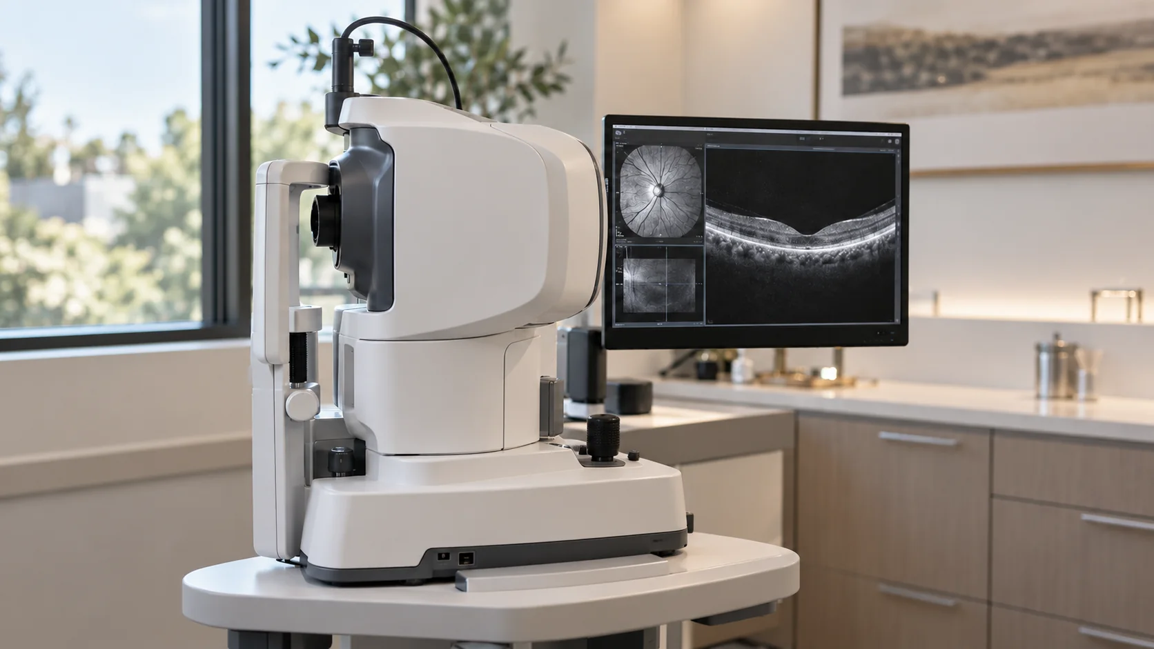
Good treatment is downstream of good measurement. The office is equipped so that decisions — is this glaucoma progressing, are these glands dying, is this cornea steepening — are made by looking directly at the structure in question.

Diagnostic imaging in use
Retina & nerve
OCT, photography, visual fields
Cornea
Topography, tomography, pachymetry
Eyelids
High-resolution meibography
Sclera
the Eaglet Eye Surface Profiler

What each tool answers

  • OCT: cross-sections of the retina and optic nerve — glaucoma, macular edema, AMD
  • Visual field testing: where peripheral vision is quietly thinning
  • Retinal photography: a baseline your future visits are compared against
  • Tonometry and pachymetry: pressure, and the corneal thickness that calibrates it
  • Corneal topography and tomography: the shape maps that stage keratoconus
  • Meibography: the structure of your eyelid oil glands
  • Scleral profilometry: the contour map a free-form scleral lens is designed from

Why baselines matter

Most eye disease is a story told in change. The first round of imaging is the chapter everything else gets compared to — which is why our optometrists in Los Angeles image before they treat.

Where it leads

Measurement drives every program here.

Common questions

Advanced diagnostics, answered.

Anything else, call the office at 310-828-2010.

  • No — they are light-based imaging and measurement. Dilation drops sting briefly and blur near vision for a few hours.

  • Glaucoma, diabetic retinopathy and early AMD are famously silent. Imaging catches them while treatment still protects vision.

  • Medically indicated testing is typically billed to medical insurance; we verify benefits when we book you.

  • A visual field test maps how much you can see to the sides, and in the center, while you look straight ahead. You keep your eye still on a central target while lights appear in different spots, and the test finds the dimmest light you can detect at each location, so missing some faint lights is normal. The map shows where side vision has been lost, which is how it helps detect glaucoma damage and track it over time. Glaucoma usually takes side vision first, and many people are unaware of it until the loss is significant, so the test can catch loss you haven't noticed.

  • Usually a few minutes per eye. In a five-center study of 126 people with glaucoma or suspected glaucoma, three versions of an automated threshold test averaged about 3, 4 and 6 minutes per eye, with only small differences in results between the quickest and the longest. Your first test becomes the baseline that later tests are compared with, since most eye disease shows itself as change over time.

Also consider

Next step

See what we can see.

Book a comprehensive exam with imaging, and keep a baseline worth comparing against.

Book Today Or call 310-828-2010