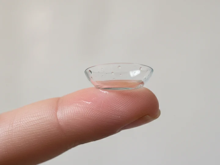
A scleral lens fitting is a short series of visits: an eye exam and a map of your eye's surface, a lens designed from that map, a visit where you learn to put the lens in and take it out, and follow-up checks that fine-tune it. A scleral lens is a large rigid contact lens that rests on the white of the eye (the sclera) and vaults over the cornea on a layer of saline. At Cohenmehr Optometry, Dr. Brandon Cohenmehr designs every scleral lens from an Eaglet Eye Surface Profiler map, so the fitting starts with measurements instead of trial and error. Here is what each step involves, and how insurance fits in.
Key takeaways
- A scleral lens fitting runs over a few visits, from the exam and eye map to the design, training and follow-up checks.
- The Eaglet Eye Surface Profiler maps the cornea and the white of the eye, and the lens is designed from that map.
- You learn to fill the lens with preservative-free saline, insert it without bubbles, remove it and clean it before you go home with it.
- Pain, redness, blurred vision, light sensitivity or a white spot on the cornea means lens out and a same day exam, or an emergency room when the office is closed.
01
Who needs a scleral lens fitting?
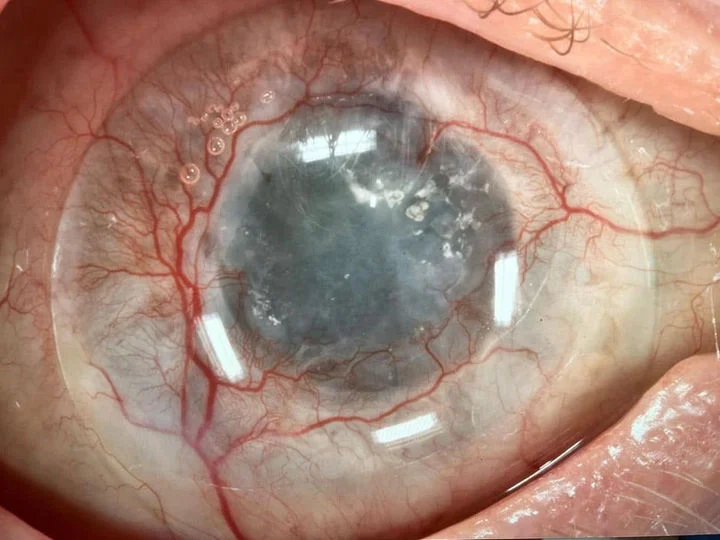
People whose vision glasses and soft lenses can't correct well, and people whose eye surface needs protection. Keratoconus is the classic reason: as the cornea bulges, an FDA review notes that spectacles may no longer adequately correct vision, and the use of rigid, scleral, or hybrid contact lenses may be needed.
A review in Clinical Ophthalmology lists the main uses as corneal ectatic diseases such as keratoconus and ocular surface conditions such as dry eyes. We also fit them after corneal transplants, after LASIK or radial keratotomy left an irregular cornea, and for eyes that can't close fully. Our scleral lens page lists the conditions we fit them for.
02
What happens at the first visit?
An exam and a map. We check your vision with your current glasses or lenses, examine the cornea and the eye's surface at the slit lamp, and measure the shape of the eye.
We also ask what you have tried: glasses, soft lenses, rigid lenses or earlier scleral lenses, how many hours a day you could wear them and what went wrong. A lens that fogged by midday, left a red ring on the white of the eye or slid off center tells us a lot about what your next design needs.
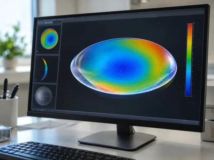
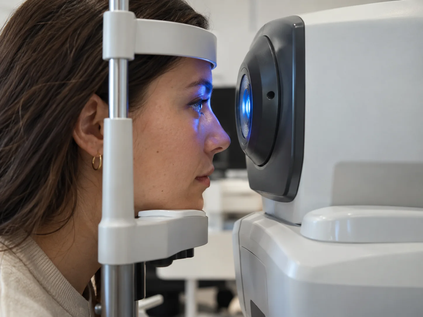
The map comes from the Eaglet Eye Surface Profiler, a camera that measures height at hundreds of thousands of points across the cornea and the white of the eye. Standard corneal topography stops at the edge of the cornea. A scleral lens lands farther out, on the sclera, so the Eaglet measures the ground the lens will rest on.
The scan takes a few seconds per eye, and nothing touches your eye. Bring your current lenses and any records from an earlier fitting, since they tell us what worked and what didn't.
03
How is the lens designed?
From your map, not from a stock lens. The lens is built on a digital model of your eye: its vault set to clear the cornea, its edge shaped to land evenly on your sclera and its optics centered over your line of sight. The Clinical Ophthalmology review describes scleral lenses as fitted to not touch the cornea, leaving a space between the cornea and the lens that fills with saline.
For eyes with an uneven sclera, we use free-form designs such as the Visionary Optics Latitude lens, whose landing zone follows the eye's measured shape in every direction. That evenness is what keeps a scleral lens steady and comfortable through the day.
04
How do you learn to insert and remove the lens?
In the office, before the lens goes home with you. Insertion and removal take practice, so the training visit is unhurried: you fill the lens with preservative-free saline, bring your face down to it, set it on the eye without trapping a bubble and remove it with a small plunger.
Saline in the bowl is for filling and rinsing only. The FDA's rule for saline is to only use sterile saline solutions for rinsing and never for cleaning or disinfecting, so you also learn the cleaning system for your lens. Our scleral lens insertion and removal guide covers each step at home, with what to do about a bubble or a lens that won't release.
05
What happens at the follow-up visits?
We check how the lens behaves after you have worn it. A scleral lens settles slightly onto the eye over the first hours of wear, so we look at it after it has been on for a while: the space it leaves over the cornea, how the edge lands on the white of the eye, your vision and your comfort.
Tell us how the days have gone. Clear vision in the morning that clouds by afternoon, redness where the edge sits or a lens that tilts when you blink are all signs the design needs a change, and they are easier to fix early than after weeks of wear.
If something needs to change, the change is made to the design against your map, and a new lens is ordered. Most fittings need one or more of these refinements. Once the lens is right, we see you for routine checks, and we look at both the lens and your eye at each one.
06
Does insurance cover the fitting and the lenses?
Often, when the lenses are medically necessary, meaning glasses can't correct your vision well enough. We bill scleral lenses to VSP and EyeMed, and to medical plans such as PPOs that come with a vision add-on. Our page on scleral lenses covered by insurance explains what the plans look at.
Plans set their own rules. EyeMed's criteria include keratoconus when vision is not correctable to 20/25 in either or both eyes using standard spectacle lenses, and VSP plan wording says a VSP network doctor receives prior approval for such cases. That is why the measurements from your first visit matter: they are what the plan reviews. Call 310-828-2010 with your insurance card and we check your benefits before the fitting.
07
When should you call us right away?
Take the lens out and call 310-828-2010 right away for a same-day exam if you have pain, redness, blurred vision, light sensitivity or a white spot on the cornea, during the fitting period or any time after. When the office is closed, go to an emergency room. The CDC's advice for these symptoms is to remove your contact lenses (if you wear them) and call your eye doctor immediately.
08
Questions patients ask
How long does a scleral lens fitting take?
The first visit, with the exam and the Eaglet map, takes longer than a routine eye exam, and the scan itself takes only seconds per eye. The full fitting runs over a few visits: the design, a training visit when the lens arrives, and follow-ups to fine-tune it. How many depends on your eye, and a free-form design made from a map usually needs fewer changes.
Does a scleral lens fitting hurt?
No. The map is taken without touching your eye, and the exam uses the same instruments as a regular eye exam. Putting a scleral lens in for the first time feels odd rather than painful, because the lens rests on the white of the eye, which is less sensitive than the cornea. Pain is not part of a normal fitting: if a lens hurts, take it out and call us.
Can I wear my scleral lenses home after the training visit?
Yes, once you can put the lens in and take it out on your own. We make sure you can do both before you leave, and you go home with the cleaning system and the saline you need. If you struggle at home, call us: a second short training visit is common.
What should I bring to a scleral lens fitting?
Your insurance card, your current glasses and lenses, and any records from an earlier fitting, such as the lens brand and specifications or old corneal maps. If you wear lenses now, ask us whether to come with them in or out, since we sometimes want to see your eye after the lens has been on for a while.
09
Sources
- Clinical Ophthalmology, 2015: Scleral lens for keratoconus
- FDA: Photrexa review summary (keratoconus and contact lenses)
- FDA: Contact lens solutions and products
- CDC: What causes contact lens-related eye infections
- EyeMed: Member FAQ (medically necessary contact lenses)
- University of California: VSP plan booklet
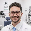
Written by
Dr. Brandon Cohenmehr, OD
Specialty contact lenses, with a focus on Ortho-K, scleral lenses, and co-managing patients with keratoconus and other corneal disease. A graduate of SUNY College of Optometry.
About Dr. Brandon →